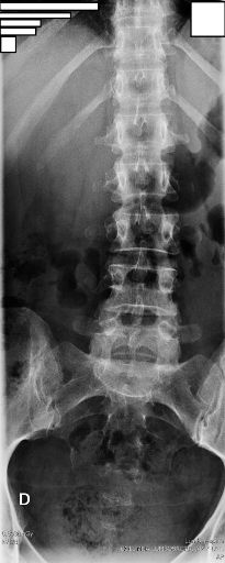
AP LUMBAR
Anteroposterior Lumbar Spine Projection • Evaluation of L1 to L5 vertebrae
Exposure Factors
High kV and mAs: Necessary to penetrate lumbar and abdominal muscle mass
Visible Anatomical Structure
The following must be clearly observed:
- T12 vertebral body (12th thoracic)
- Heads of 11th and 12th ribs (floating ribs)
- Vertebral bodies L1 to L5
- Part of the sacrum
- Sacroiliac joints
- Intervertebral disc spaces
- L5-S1 joint (lumbosacral)
- Spinous processes
- Transverse processes
- Pedicles
Cassette Sizes and Centering
STANDARD CASSETTE
Orientation: Portrait
Cassette Centering: 4th lumbar vertebra (L4)
Central Ray: 4th lumbar vertebra (L4)
Recommended
RARELY USED CASSETTE
Orientation: Portrait
Cassette Centering: 3rd lumbar vertebra (L3)
Central Ray: 3rd lumbar vertebra (L3)
Seldom used
Both in portrait orientation - Centering varies by cassette size
Patient Positioning
Supine Position (Preferred)
Alternative: Upright Position
If the patient cannot lie flat on the table:
- Perform standing at the wall bucky
- Same alignment and centering criteria apply
- Useful for patients with acute pain or mobility limitations
- Allows for weight-bearing evaluation (functional position)
Central Ray Point
Important: Cassette and central ray centering must match based on the size used
Optimal Image Characteristics
Vertebrae L1-L5
All included in field
Disc Spaces
Intervertebral symmetry
Symmetry
Centered spinous processes
L5-S1 Joint
Clearly visible
Ribs 11-12
Heads visible
SI Joints
Symmetrical sacroiliac
Common Technical Challenges
Frequent issues in AP lumbar projections:
- Patient rotation causing spinous process asymmetry
- Excessive lordosis due to un-flexed legs
- Intestinal gas superimposition obscuring vertebrae
- Incomplete field missing T12 or sacrum
- Insufficient exposure in obese patients (low kV/mAs)
- Breathing during exposure causing blurriness
- Incorrect centering based on cassette size
Solution: Flex legs, instruct apnea, adjust kV/mAs by biotype, verify centering by size
Special Considerations
Obese Patients
Increase kV up to 90-100 and mAs. Consider grid-less technique if necessary.
Geriatric Patients
Possible osteoporosis requires reduced kV. Use extra padding for comfort.
Acute Pain
Upright position might be better tolerated. Use support cushions.
Scoliosis
Align based on the primary curve. Multiple exposures may be required.
Bowel Preparation (Optional)
To reduce intestinal gas superimposition:
- Fasting 4-6 hours before the study
- Bowel evacuation prior if possible
- Abdominal compression with band during exposure
- Expose on expiration to decrease abdominal volume
These measures improve L4 and L5 vertebrae visualization
Patient Instructions
"You must not breathe during the exposure"
Full Sequence:
1. "Flex your knees to flatten your back against the table"
2. "Stay completely straight"
3. "Do not tilt your body to either side"
4. "Take a deep breath in, then blow all the air out"
5. "Hold your breath and do not move"
6. "Relax when I tell you"